How Does The Brain Deal With Visual Changes Following Cataract Surgery?
Cataracts are the leading cause of blindness worldwide. However, there is a simple surgical procedure that can remedy this problem and restore sight to patients. However, this cataract operation involves a series of changes that are interesting to know.
Eye cataracts are a fairly common visual problem. In fact, according to several studies, over 60% of the world’s population over 75 years of age develop some form of cataract. Cataracts therefore mainly affect older people.
In addition, the symptoms of this ocular pathology are well known. Patients suffer from a progressive degradation of the functionality of vision which can become disabling.
What is cataract?
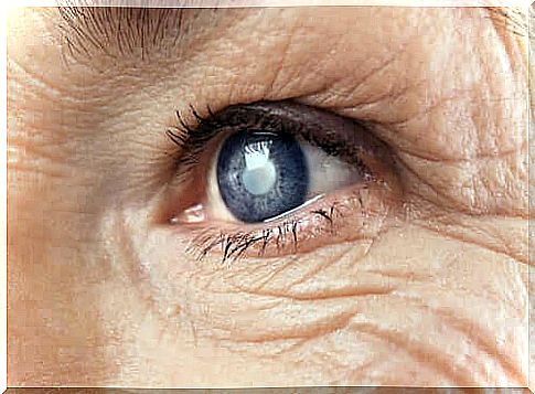
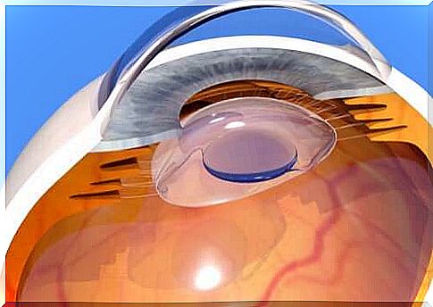
What is meant by ocular cataract is nothing more than the clouding of the lens. The lens is the transparent lens of the eye, through which the eye can focus. It is located behind the iris and the pupil.
When cataracts appear, this lens gradually becomes cloudy. This is due to an accumulation of epithelial cells that change color for pathological reasons.
These cells reproduce and then obstruct the normal passage of light through the lens. Indeed, their translucent characteristics and their brown coloring represent a physical barrier to the passage of light. It’s like looking at the world through a foggy windshield. In this way, vision is affected.

There is also another clinical case. This is the secondary cataract. This ocular pathology is in fact very similar to the previous one. It occurs after the surgical removal of the cataract when residues of this dark epithelium reproduce again, making the structures of the eye opaque again.
The resulting symptomatology is similar to that of primary cataract. However, the eye damage is lighter and less extensive. Thus, it is sufficient to remove this layer of tissue by laser to correct the problem. It is a quick, painless and risk-free procedure.
Symptoms of cataracts
The formation of cataracts is progressive and it is more or less quickly depending on the type of cataract in question. In all cases, the person concerned feels the following visual discomfort:
- Decreased visual acuity
- Photophobia and glare
- An alteration of chromatic perception: that is, the colors lose their intensity and fade.
- An alteration in the perception of the surrounding space
And, in general, any kind of visual change that can result from the fact that light reflected from objects cannot enter the eye properly. And, even if it does, visual perception remains dark and distorted.
Also, to add to the confusion, when cataracts develop, varying degrees of pupil dilation or contraction can mask or exacerbate symptoms.
Thus, if the pupil is very dilated, much of the light entering the eye will “dodge” the cataract and vision will be relatively close to normal. However, if the pupil is very tight – that is, in low light environments – the cataract will block more light and vision will be severely impaired.
Causes of eye cataracts
Cataracts are generally part of the body’s normal aging process. In this sense, the development of cataracts is a logical consequence of advanced age. However, not all people who reach a certain age have this condition.
In a number of cases, however, the cause is genetic. In this case, it is called a congenital cataract. There is thus a small percentage of babies who are born with this problem. If so, then surgery is recommended for the child and the results are generally good.
Without speaking of idiopathic cataracts, that is to say of no known origin, there are three other possible origins of this ocular pathology:
- A high degree of myopia which then influences the ocular morphology
- The presence of eye trauma
- Prolonged pharmacological treatments, in particular based on steroids and in particular corticosteroids. This is for example the case of chronic cortisone intake for rheumatism or antiallergic drugs in people with allergies
In all cases and whatever the cause of the cataract, the consequences are such on the daily performance of the patient that they generally lead to a whole series of psychological inconveniences. Among them anxiety and depression.
What does cataract surgery involve?
Cataract surgery is now considered a simple and safe surgical procedure. It is even an outpatient procedure. The cataract operation should therefore not frighten you. Every day, hundreds of such operations are performed around the world and the success rate is very good.
To put it simply, the operation involves physically extracting the cataract using instruments inserted into the anterior part of the eyeball.
Since the tissue to be removed touches the healthy eye tissue, in other words the lens, it is necessary to remove part or all of the lens. The eye then becomes aphakic, that is to say deprived of its crystalline lens. It loses its own ability to focus. Let us remember that it is this same crystalline lens, our natural lens, which allows the focusing and, consequently, which allows the accommodation of the sight.
Intraocular lenses
To overcome this structural and functional deficiency, an Intraocular Lens (IOL) is implanted to replace the lens. This then makes it possible to focus the images again. And this, with characteristics complementary to those of the IOL of the opposite eye.
Indeed, cataracts are almost always bilateral, that is to say, it manifests in both eyes at the same time. Each eye forms light projections that focus on the retina. The combination of these two pieces of information will then give rise to a visual representation, at the level of the brain.
But as so often, artifices hardly replace the natural. Intraocular lenses are indeed artificial lenses that have a given shape. They therefore do not have the ability of the lens to change shape to focus. This therefore dramatically changes the way the visual system operates at different levels.
The brain is the organ responsible for building the final perceptual image. Following a cataract operation, he must therefore readjust to this new way of interpreting information coming from the eyes. This is why people who have such surgery may need some time for this readjustment to take place. It can take up to a month. During this time, their vision lacks precision and they experience some visual discomfort.
Thus, thanks to the plasticity of the brain and the ability of our neural systems to reorganize and adapt, this adjustment process is made possible so that the patient regains good visual capacity.
Here is an overview of the elements of the visual process that can be disturbed after cataract surgery. These are alterations that the brain will learn to correct in order to regain the clearest possible vision.
The changes the brain must learn to accommodate
A continuous effort of concentration and the disappearance of the time required to focus the view
As we said earlier, IOLs only focus and do not have the ability to adjust focus. Thus, as long as the eyes are open, one can see accurately at any distance. Whether near or far. And this without having to force the eyes or wait a few thousandths of a second for the gaze to focus on another point.
This situation causes a strange sensory sensation at first. However, over time you get used to this distortion.
After cataract surgery: you need more light to see well
People who suffer from cataracts tend to avoid places that are too bright. Indeed, the light dilates the pupil and causes visual discomfort in them. On the other hand, following a cataract operation and the implantation of intraocular implants, they seek the light! Thus, the brighter the environment, the better their focus and vision.
This is mainly due to the fact that the artificial lens does not cover the entire diameter of the pupil when the latter is very dilated. If this happens, some of the light enters the eye from outside the IOL. This then causes double or blurred vision and light halos.
A need to move in order to orient oneself in space
Since IOLs focus in a fixed and determined manner, patients with cataract surgery often find the need to move and place their head at different angles in order to adjust their vision to perceive objects that are at a specific distance. . Indeed, they have lost their ability to adapt their lens.
After cataract surgery: residual presbyopia
In general, IOLs are designed to focus on objects that are at medium to long distances. As a result, people with cataract surgery find it more difficult to see at short distances. Patients sometimes need to use a magnifying glass to see them up close.
Visual changes under the effect of physical stress
Intraocular pressure is defined as the pressure exerted by liquid substances on the eye. IOLs have a lower degree of fixation in the eye than natural lenses. Therefore, changes in intraocular pressure that result from physical stress produce slight transient movements of the IOL. This phenomenon causes distortions in the observed image.
After cataract surgery: a tendency to have sore eyes
The ocular or connective epithelium is damaged during cataract surgery. It then develops microscopic scars that make the eye more sensitive to external elements. For example, smoke, dust, etc.
Different visual convergences
It is the brain that builds a final image. This is the result of merging two images of the same object. Each image from each eye.
Because intraocular implants are placed a little differently from the original lens, the exact point in the retina where they project light is also slightly different.
Thus, the two images to be merged to form the final representation come from slightly different areas. The brain must therefore relearn how to perform this fusion correctly.
After a cataract operation: a distortion of the visual plan and the dimensions of the objects
IOLs implanted after cataract surgery usually produce a flattened perception of the visual field. For this reason, the objects then appear as having less relief.
Also, these same IOLs tend to overwrite the image they are focusing on. As a result, the eye operated on for cataracts perceives the observed objects as smaller than they were previously. The brain will also have to get used to this to get rid of the resulting visual discomfort.
Presence of visual artifacts
In medicine, an artefact is defined by the presence of an element that should not be in a certain place. In the field of ophthalmology, a visual artifact is something that is perceived by the visual system but which does not actually exist.
People who have IOLs implanted can thus, in some cases, perceive certain pieces of the lens itself or even the movement of the lens.
This situation is uncomfortable and disconcerting. It often distracts the patient’s attention. Again, thanks to neuroplasticity and adaptation to the new visual environment, this problem quickly becomes a minor problem.
To conclude, whether you have already had a cataract operation or need it at some point, the few points mentioned in this article are important to know in order to explain some of the visual phenomena that result from the cataract operation. . It will also allow those about to be operated on to personally prepare for these changes.

The vision of color is a subject as interesting as it is complex. Many wonder if we really all perceive the color of the same my …